
CSBIO

What Is iPSC Differentiation?

What is iPSC differentiation, and why does it matter? Induced pluripotent stem cells, or iPSCs, are adult cells reprogrammed into a flexible, stem-cell-like state. Differentiation guides them toward specialized cell types, such as neurons, heart muscle cells, or insulin-producing cells. In the lab, this change unfolds through carefully timed signals, media changes, and checks under the microscope. It is not a simple switch.

Nobel laureate Shinya Yamanaka, whose work helped establish iPSC reprogramming, once said, “I could not stop thinking about the patients.” His words capture an important motivation, but promise is not proof. Researchers use iPSC differentiation to model disease, study development, and investigate potential treatments. Results depend on the starting cell line, culture conditions, and how carefully each cell type is verified. A dish of beating cells may look convincing. It still needs rigorous testing.

This guide explains the process, common methods, and practical challenges behind iPSC differentiation. It also considers cell identity, purity, and reproducibility—details that can shape whether an experiment is useful. Some protocols work beautifully in one laboratory and falter in another. That is worth admitting. A clear understanding of the method helps readers judge what the results show, and what they do not.
What Is iPSC Differentiation?

How iPSCs Acquire Specialized Cell Identities

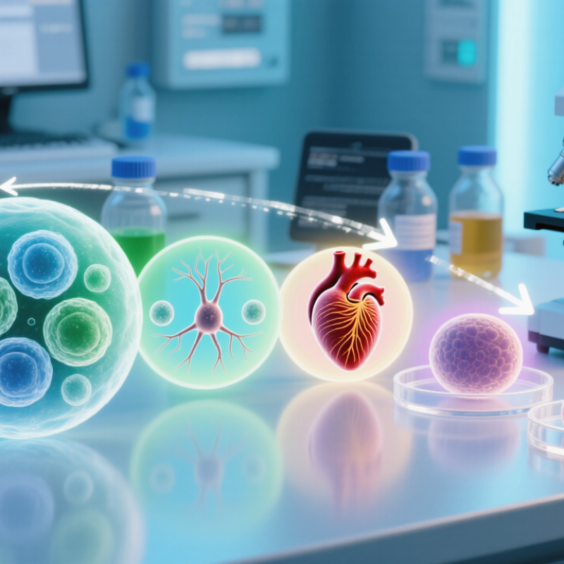
Induced pluripotent stem cells (iPSCs) can be guided toward specialized identities by recreating signals involved in early development. Differentiation is not a single leap; cells pass through intermediate states as gene activity and behavior change. Researchers adjust growth factors, culture media, and timing to steer pathways linked with a target tissue. A timed sequence matters: too much signal, or a missed media change, may alter the outcome. In culture dishes, emerging cells may change shape, form clusters, and begin expressing characteristic markers. Not a switch.

Appearance alone cannot confirm a cell’s identity. Researchers also examine marker patterns and test whether cells perform expected functions, such as contracting or producing a specific substance. Results can vary between batches, and some cells may remain immature or take an unexpected path. The process is less tidy than diagrams suggest. Careful controls and repeated measurements help distinguish genuine specialization from look-alike changes, though no single test answers every question. Researchers must also consider how culture conditions affect the cells being measured, since a promising marker profile does not guarantee stable function.

Key Signals That Guide iPSC Differentiation

What Is iPSC Differentiation?

Key Signals That Guide iPSC Differentiation

Induced pluripotent stem cells, or iPSCs, can become specialized cells when exposed to carefully timed developmental cues. The process resembles early embryonic development, but it is guided in controlled laboratory conditions. Signals do not act alone. Their effects depend on dose, timing, cell density, and the cells’ starting state.

Activin and Nodal signaling can support mesendoderm formation, while BMP and Wnt activity may steer cells toward particular developmental paths. FGF signals help regulate growth and lineage progression. A brief Wnt pulse, for example, may encourage cardiac mesoderm, while prolonged exposure can change the outcome. Small shifts matter. The same molecule can have different effects at different stages.

Researchers adjust these cues in sequence, often changing growth factors as cells develop. They also monitor cell shape and use molecular markers to check whether the intended lineage is emerging. A dense patch of cells may respond differently from a sparse one, even in the same medium. There is no universal recipe. Protocols can vary between cell lines, and a promising marker does not prove that every cell has matured properly. That uncertainty deserves attention, especially when interpreting results across experiments.

Common Strategies for Directing Cell Development

iPSC differentiation guides induced pluripotent stem cells toward specialized cell types, such as neurons or heart muscle cells. The central challenge is recreating the signals cells encounter during development. Researchers often use growth factors or small molecules to activate or block specific pathways. Timing matters. A signal given too early can produce a different outcome than the same signal given later.

Protocols usually apply cues in stages, with each stage supporting a change in cell identity. For example, researchers may first encourage cells to form an early precursor, then provide signals that steer it toward a target tissue. The culture surface also matters: its stiffness and coating can influence how cells attach and develop. Teams monitor cell shape and use molecular markers to check whether the intended lineage is emerging. They may adjust signal concentrations, feeding schedules, or cell density when results vary. It is easy to overread a promising marker; no single measurement proves that cells are fully mature or functional. Even a carefully planned protocol can behave unevenly across batches. That variability deserves attention, not concealment.

How Differentiated Cells Are Identified and Assessed

What Is iPSC Differentiation?
How Differentiated Cells Are Identified and Assessed

A dish of differentiated cells may look uniform, but appearance alone cannot confirm cell identity. Researchers assess several kinds of evidence. Microscopy can reveal changes in shape, such as elongated cells or organized clusters. These features are useful clues, not proof. Looks can deceive.

Marker testing provides stronger evidence. Immunostaining can show whether cells carry proteins linked to a target lineage, while flow cytometry estimates how many cells express selected markers. Gene-expression tests offer another view. A reliable assessment uses a panel of markers, including markers that should be absent, rather than relying on one result. Controls matter: undifferentiated iPSCs and appropriate comparison samples help reveal misleading signals.

Function is also important. Neuron-like cells, for example, can be assessed for electrical activity; other cell types may be tested for characteristic secretions or metabolic behavior. Researchers also track cell survival, purity, and any remaining pluripotent cells, since these can affect how results are interpreted. Measurements at more than one time point can expose unstable differentiation. Still, no single assay settles every question, and results may vary between batches. A tidy data set can hide that variation. It is worth asking whether the tests support the intended use, not merely whether the cells appear convincing.

What Is iPSC Differentiation? — How Differentiated Cells Are Identified and Assessed
Differentiated cell type Common identity markers Ways to assess identity Evidence of function Important interpretation notes
Neurons
Derived from the ectodermal lineage
MAP2 and TUBB3 (βIII-tubulin) are commonly used neuronal markers. PAX6 and SOX1 can indicate neural progenitor stages. Immunostaining or flow cytometry for protein markers; gene-expression analysis; microscopy to examine neuronal morphology. Electrophysiology can test action potentials and synaptic activity. Calcium imaging can assess stimulus-related activity. Neuronal cultures may contain progenitors or multiple neuronal subtypes. Marker expression alone does not establish mature neuronal function.
Cardiomyocytes
Derived from the mesodermal lineage
TNNT2 (cardiac troponin T), ACTN2 (α-actinin), and NKX2-5 are commonly assessed. Immunostaining, flow cytometry, or gene-expression analysis; sarcomere organization can be examined by microscopy. Assess spontaneous contraction, calcium transients, and electrical activity. Patch-clamp or multielectrode-array measurements can provide electrophysiological evidence. Beating is useful but does not by itself prove a mature cardiac phenotype. Cell maturity and subtype composition can vary between differentiation batches.
Hepatocyte-like cells
Derived from the endodermal lineage
HNF4A and albumin (ALB) are commonly assessed; additional markers may include alpha-1 antitrypsin (SERPINA1). Immunostaining or gene-expression analysis, combined with measurement of secreted albumin. Measure albumin secretion and activities of drug-metabolizing enzymes, such as CYP3A4, using appropriate functional assays. iPSC-derived hepatocyte-like cells may retain features of fetal or immature cells. A single marker or assay is not sufficient to establish adult-like function.
Endothelial cells
Derived from the mesodermal lineage
CDH5 (VE-cadherin), PECAM1 (CD31), and VWF are commonly used endothelial markers. Flow cytometry or immunostaining for multiple markers; microscopy can assess cell-junction patterns. Barrier-permeability measurements and assays of responses to angiogenic signals can provide functional evidence. Some markers are also found in other cell populations. Use a combination of identity markers and functional tests.
Pancreatic beta-like cells
Derived from the endodermal lineage
INS (insulin), C-peptide, PDX1, and NKX6.1 are commonly assessed. Immunostaining or gene-expression analysis; intracellular insulin and C-peptide can be measured using validated assays. Glucose-stimulated insulin secretion can test whether secretion changes in response to glucose. Measuring C-peptide helps distinguish newly produced insulin from insulin taken up from the culture medium. Insulin expression alone does not confirm glucose-responsive beta-cell function. Assess secretion under controlled conditions and include appropriate controls.
Across differentiated cell types
Culture purity and quality
Assess lineage-specific markers alongside markers of undifferentiated pluripotent cells, such as OCT4 (POU5F1) and NANOG. Use complementary approaches such as flow cytometry, immunostaining, and gene-expression analysis. Record the fraction of cells positive for relevant markers. Compare functional readouts across replicates and differentiation batches, using defined assay conditions and positive or negative controls where appropriate. There is no single universal marker panel or acceptance threshold for every cell type. Marker selection and cutoffs should be defined for the intended research use.

In brief: iPSC differentiation is the process of directing induced pluripotent stem cells toward a specialized cell type. Identity is best assessed using multiple lineage markers together with relevant functional tests, rather than relying on one marker alone.

Research Uses and Challenges of iPSC-Derived Cells

What Is iPSC Differentiation?

Research Uses and Challenges of iPSC-Derived Cells

Induced pluripotent stem cells can be guided toward specialized cell types, including neurons, heart muscle cells, and liver-like cells. Researchers use these cells to model diseases in laboratory dishes, where they can observe changes such as altered electrical activity or protein buildup. A patient-derived cell line may help reveal how a particular genetic variant behaves. It does not, by itself, prove what will happen in a person.

These cells also support early drug research. Scientists can compare treated and untreated cultures, measure cell survival, and track responses across repeated experiments. Some derived heart cells contract visibly under a microscope. Useful, but not identical to adult heart tissue. Many iPSC-derived cells remain developmentally immature, and cultures can contain a mix of cell types. Differentiation protocols may also produce different results between batches.

Careful quality checks matter. Researchers often assess cell identity, purity, function, and genetic stability before drawing conclusions. Even then, a clean marker profile may not guarantee normal behavior. Residual undifferentiated cells can raise safety concerns, while immune compatibility is not automatic, even when cells come from a patient. Scaling production consistently remains difficult.

It is worth being cautious: promising results in a dish are a starting point, not evidence of a proven treatment.

Close Menu